Understanding Your Skin Cancer Detection and Treatment
Depending on the type and stage of skin cancer, your healthcare provider may suggest a range of options. This page discusses some of the most common treatments, including topical therapies, electrosurgery, curettage, and Mohs surgery. This page also showcases the most common detection methods, as well as some of the best non-invasive detection methods that are often not utilized if a patient isn't aware of their options.
Non-Invasive Detection Methods
Reflectance Confocal Microscopy (RCM)
- This is essentially a high-powered "living microscope" that allows doctors to see your skin cells in real-time while they are still on your body.
- Instead of cutting out a piece of skin and sending it to a lab, the doctor uses a special laser to shine a light into the skin.
- This light reflects off your cells and sends a sharp, black-and-white image to a computer screen, showing individual cells and structures as if they were under a traditional microscope.
- It is incredibly accurate for spotting Melanoma and Basal Cell Carcinoma because it can see the specific shape and arrangement of the cells.
- While it only looks at the very top layers of the skin, it is often used as a "second opinion" for a suspicious mole; if the cells look healthy through the RCM, the doctor might be able to save you from getting a physical biopsy and a scar.

Image Credit: Kris Hanning / University of Arizona Health Sciences via Photonics Media.
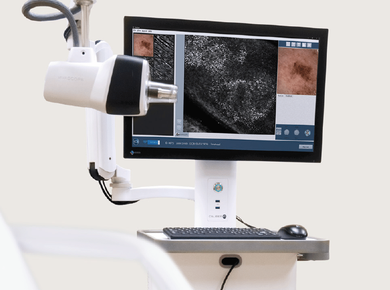
Image Credit: The Skin Hospital (Australia)
Optical Coherence Tomography (OCT)
- Essentially a high-tech "camera" that uses light to see through your skin without any cutting or needles. Instead of taking a picture of just the surface, it sends light beams deep into the skin and measures how they bounce back, almost like how a submarine uses sonar to find things underwater.
- This creates a 3D, 2-millimeter-deep map that shows the different layers of your skin in real time on a screen. It is incredibly helpful for finding Basal Cell and Squamous Cell cancers because doctors can see the "roots" or nests of the tumor that are invisible to the naked eye.
- While it’s great for those types, it isn't used as much for Melanoma yet, because the dark pigment in those spots blocks the light from seeing clearly.

Image from Al-Arashi & Wang, "Deep Learning in the Detection and Diagnosis of Melanoma," Current Oncology (2023). Licensed under CC BY 4.0.
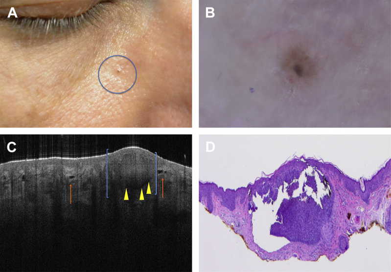
Image from Rajadhyaksha et al., "Reflectance Confocal Microscopy of Skin In Vivo," Dermatologic Clinics (2017).
Invasive Detection Methods
Sentinel Lymph Node Biopsy
- A surgical procedure to determine if cancer cells have spread to the lymphatic system by testing the "first" drainage node.
- Commonality: Regularly used for cancers that have reached a certain thickness or depth.
- Used For: Primarily Melanoma and occasionally high-risk Squamous Cell Carcinoma or Merkel Cell Carcinoma.

Image Credit: Cleveland Clinic Health Library

Image Credit: Dr. Finbar McGrady, Dr. Finbar’s Skin Clinic (2024).
Shave, Punch and Excisional Biopsies
Shave Biopsy
- A small blade is used to remove the outermost layers of the skin (the epidermis and a portion of the dermis).
- When It’s Used: Most common for suspected Basal Cell or Squamous Cell Carcinomas, or lesions that are primarily on the surface.
- Patient Benefit: Quick, typically requires no stitches, and heals similarly to a small graze or scrape.
Punch Biopsy
- A circular tool (like a tiny cookie cutter) is used to remove a deeper "core" of skin tissue.
- When It’s Used: Vital for looking "beyond the surface" when a provider needs to check the full thickness of a lesion or rash.
- Patient Benefit: Provides a highly accurate cross-section of the skin layers, ensuring a more definitive diagnosis for deeper growths.
Excisional Biopsy
- The entire suspicious area or tumor is removed, often along with a small margin of healthy skin.
- When It’s Used: The gold standard for suspected Melanoma to ensure the entire lesion is captured for laboratory staging.
- Patient Benefit: Often serves a dual purpose—it acts as both the diagnostic test and the first step of treatment by removing the growth entirely.
Less Invasive Removal Methods
Cryosurgery
- Using liquid nitrogen to freeze and destroy the cancerous tissue.
- Commonality: Common for pre-cancers, but less common for confirmed invasive cancers.
- Used For: Pre-cancerous Actinic Keratoses and very thin, superficial BCC or SCC.

Image Credit: Northboro Medical Center; Terese Winslow
Topical Chemotherapy or Immunotherapy Creams
- Medicated creams applied at home over several weeks to destroy cancer cells through the skin surface.
- Commonality: Fairly common for patients who want to avoid surgery for very thin, early spots. Most commonly prescribed is called Fluorouracil.
- Used For: Superficial BCC, early SCC (Bowen’s disease), and pre-cancerous Actinic Keratoses.

Image Credit: Verywell Health
Immunotherapy
- A systemic drug treatment (often IV) that boosts the body's immune system to recognize and fight cancer cells.
- Commonality: Common for advanced stages; has become the primary treatment for cancers that have spread.
- Used For: Advanced Melanoma, Merkel Cell Carcinoma, and advanced SCC that cannot be treated with surgery or radiation.

Image Credit: DTU Health Tech / Technical University of Denmark
Invasive Removal Methods
Curettage and Electrodesiccation (Scrape and Burn)
- A sharp tool (curette) scrapes away the tumor, followed by an electric needle to cauterize the base and kill remaining cells.
- Commonality: Very common for low-risk, surface-level spots on the trunk or limbs.
- Used For: Superficial BCC and early SCC. It is almost never used for Melanoma.

Image from Thompson & Driscoll, "Skin Cancer: Prevention and Management," Obstetrics and Gynecology Clinics (2011).
Wide Local Excision
- A more invasive version of standard excision that removes a larger "clear zone" of tissue to ensure aggressive cells are gone.
- Commonality: Common and standard for any confirmed invasive cancer.
- Used For: Specifically used for Melanoma, Merkel Cell Carcinoma, and Atypical Fibroxanthoma (AFX).

Image Credit: Storwick Dermatology
Mohs Micrographic Surgery
- The surgeon removes thin layers of skin one at a time, checking each under a microscope until only cancer-free tissue remains.
- Commonality: Extremely common; the gold standard for areas where preserving skin is vital (face, ears, hands).
- Used For: Basal Cell Carcinoma (BCC), Squamous Cell Carcinoma (SCC), and sometimes early-stage Melanoma (Melanoma in situ). It is also the preferred treatment for rare tumors like Dermatofibrosarcoma Protuberans (DFSP).

Image Credit: NIH / National Cancer Institute
Standard Surgical Excision
- The doctor cuts out the visible tumor along with a small border of healthy skin and stitches the wound closed.
- Commonality: The most common surgical treatment for almost all skin cancer types.
- Used For: BCC, SCC, Melanoma, and rarer types like Merkel Cell Carcinoma or Sebaceous Carcinoma.

Image Credit: MyPathologyReport.ca
Radiation Therapy
- Using high-energy beams to kill cancer cells, often over multiple sessions.
- Commonality: Moderately common; usually a "backup" or used for patients who aren't candidates for surgery.
- Used For: BCC, SCC, and Merkel Cell Carcinoma. It is less commonly used for Melanoma except in specific palliative or advanced cases.

Image Credit: Water's Edge Dermatology
When it comes to skin cancer treatment, it's essential to discuss all available options with a qualified healthcare provider. Each treatment has its own benefits and risks, and the best approach will depend on the specific circumstances of your diagnosis. Staying informed and proactive about your skin health can make all the difference in successful treatment outcomes.